Neurodegeneration is a complex process, and at its core, it involves a series of triggers that gradually erode the structure and function of neurons, leading to a cascade of debilitating effects. Neurodegeneration manifestation includes progressive cognitive decline, which often manifests as a gradual decline in cognitive functions, affecting memory and other cognitive abilities. In Parkinson’s Disease and Amyotrophic Lateral Sclerosis, neuronal degeneration leads to motor dysfunction, muscle weakness, and tremor. The common denominator of Neurodegeneration is the progressive loss of neurons, which can result in brain atrophy and functional deficits.
For 45 years, Enzo has been collaborating with researchers around the world, developing unique solutions to advance neurodegeneration research and accelerate next-generation therapies’ development.
With our comprehensive catalog of unique products, we are committed to providing the tools and support you need and driving innovation and advancement in neuroscience.
Biomarkers for neurodegenerative diseases are crucial as diagnostic and prognostic tools and to effectively monitor the patient treatment response after disease-modifying therapies.
Trauma, infection, stroke, toxic metabolites, or autoimmunity can cause inflammation in the nervous system. Repeated or chronic inflammation promotes neurodegeneration.
Accumulation of pathological proteins and abnormal protein aggregates, such as plaques and tangles are common features of several neurodegenerative diseases.
Inflammation
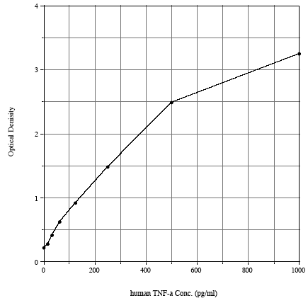
Trauma, infection, stroke, toxic metabolites, or autoimmunity can cause inflammation in the nervous system. Repeated or chronic inflammation leads to cellular neuroinflammation. Pro-inflammatory cytokines such as TNF-α , IL-1β, and IL-6 IFN-γ play a significant role in neurogenerative disorders. For instance, TNF-α, a pro-inflammatory cytokine, acts as a modulator in the acute phase of inflammation, triggering inflammatory cytokine signaling cascades.
Several mitochondrial dynamic imbalances have been associated with neurodegeneration. Misfolded protein aggregates can affect organelle homeostasis. Mitochondria accumulation of toxic level of calcium contribute to the dysregulation of homeostasis and releases apoptotic factor that further contributes to cytotoxicity.
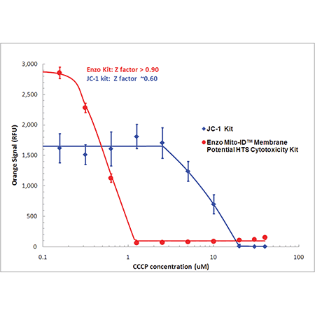
Detect mitochondrial perturbations with 10 times more sensitivity than JC-1. Mitochondrial membrane potential (MMP) was evaluated in HeLa cells treated with CCCP using MITO-ID® dye (red) or JC-1 (blue). Using a conventional fluorescence microplate reader, MMP was shown to decrease with increasing CCCP concentration as indicated by a decrease in orange fluorescence. Improved aqueous solubility of the dye and no-wash protocol minimizes variability, leading to a higher Z-factor (> 0.9) than that obtained with JC-1.
Aberrant Proteostasis
A subclass of neurodegenerative diseases is associated with dysregulation of proteins homeostasis or proteostasis. When cells cannot efficiently clear or degrade misfolded proteins, they aggregate and assemble in a dynamic and specialized structure called aggresome. The aggresome is a protective mechanism within the cells to sequester and concentrate these aggregated proteins, thereby reducing their detrimental effects.
Quantitatively detecting misfolded proteins and inclusion bodies is essential in understanding neuronal cell degeneration. PROTEOSTAT® Aggresome Detection Kit has been a valuable tool for studying aggresome-related neurodegenerative diseases in cell-based and animal models. This assay has been highly validated in several research papers.
Increased sensitivity versus alternative dyes (Thioflavin T) allowing analysis by fluorescence microscopy and flow cytometry of a variety of samples (cells and tissues)
Reliable and simple assay does not require protein mutations or genetically engineered cell lines
Validated under a wide range of conditions, from neurogenerative disease studies to oxidative stress research
Fix-and-perm assay allowing co-staining of PROTEOSTAT® with antibodies against a variety of targets such as protein aggregates, protein degradation machinery, etc.
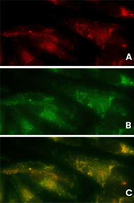
Detection of aggresomes in HeLa cells, treated with proteosome inhibitor MG-132 for 12 hours with (A) PROTEOSTAT® Aggresome Detection Kit (ENZ-51035) (red), (B) aggresome marker p62 antibody conjugated with fluorescein (green). (C) Composite image shows co-localization of aggresomes and p62 (yellow).
Cell Death
Cell death is one of the main factors leading to neuronal loss in neurodegenerative diseases. Neuronal cell death can be triggered by immune cells during inflammation, extreme impairment of mitochondria, overloaded autophagy pathways, or structural damage due to protein aggregates.
CYTO-ID® Autophagy Detection Kit 2.0 (ENZ-KIT175) used to detect autophagy in HeLa cells cultured in (A) media under normal conditions, (B) starvation media (EBSS) treated with 40uM Chloroquine for 4 hours. Starved cells show a higher quantity of autophagic vacuoles compared to cells under normal conditions (fluorescent green).
Epigenetic Alterations
Changes in epigenetic regulation, such as altered DNA methylation, histone post-translational modification, and chromatin remodeling, maybe involved in the development of several neurodegenerative diseases, including Alzheimer’s and Huntington’s disease.
A rapid, reliable method to isolate histone proteins in mammalian cells and tissues for histone modification applications.
EPIXTRACT® Total Histone Extraction Kit provides a simple and reliable method to isolate histone proteins in 1 hour. Extracts can be used for various downstream applications, such as post-translational modification assays.
High Efficiency - extract histones from as low as 105cells or 1mg tissue
Simple - optimized protocol with easy steps
Reliable - reproducible results that keep post-translational modifications intact
Histone extracts were prepared from MCF-7 cells using the EPIXTRACT® Total Histone Extraction Kit and acetyl histone H3-K9 was quantified
Biomarkers
Cell death is one of the main factors leading to neuronal loss in neurodegenerative diseases. Neuronal cell death can be triggeredx by immune cells during inflammation, extreme impairment of mitochondria, overloaded autophagy pathways, or structural damage due to protein aggregates.
AMPIVIEW™ RNA probes are uniquely designed with the precision of targeted, sequence-specific RNA probes powered by Enzo’s LoopRNA ISH™ technology to deliver superior sensitivity. ISH results with AMPIVIEW™ RNA probes preserve sample morphology, and the signal of the targeted biomarker can be seen under the light microscope.
Detection of unique nucleic acid targets (DNA/RNA) down to a single cell level
High sensitivity and reliability
Flexible products ready for any workflow (Manual or Automated)
The Enzo portfolio of neuroscience antibodies includes hundreds of monoclonal and polyclonal antibodies validated for immunohistochemistry(IHC) or immunocytochemistry (ICC) protocols. Major targets include markers of neurodegenerative disease, ion channels, neurotransmitters and their receptors, neurofilament and cytoskeletal targets, synaptic vesicle markers, and more. Each of our antibodies is backed by our Worry-free Antibody Trial Program.
Enzo has first-hand experience with unique research challenges. As an integrated biotechnology and life sciences company, we take pride in delivering dependable products and services that advance your neuroscience research from discovery to clinical trials. Our extensive experience manufacturing over 20,000 life science products and platforms gives us the tools to create complete solutions from genomic analysis, protein analysis, cellular analysis, tissue analysis to small molecule chemistry.
Rely on 45 years of innovation, technology development, and manufacturing know-how to support your neuroscience discovery, no matter how small or complex they may be. Our comprehensive Life Sciences Contract Services supports the development of customizable, unique, and efficient solutions for your neuroscience research.
As our understanding of neurodegenerative disease origins increases, so does the need for innovative, high quality research tools for developing potential therapeutics. The breadth of Enzo’s expertise across research disciplines from proteostasis and cell analysis, to small molecule chemistry and assay development provides a unique collection of innovative reagents and assays to assess the impact of neurodegeneration on critical neural signaling networks.
Neuroactive Peptide & Stress Hormone ELISAs
Put our expertise in building ultrasensitive, high-specificity ELISAs for small molecule detection to work for you!
The most sensitive colorimetric ELISA kits for detection of cAMP and cGMP, key second messenger signaling molecules
FLUOFORTE® Calcium Assay Kits
Brighter, More Robust Fluorescent Calcium Mobilization Assays
Neuromodulating Compounds
Focused Compound Libraries for Neuromodulator Screening
Tired of procuring small molecules from multiple sources?
Enzo has a long and successful history of identifying, synthesizing, and commercializing known bioactives for use as research tool compounds and assembling focused sets of compounds for chemical genomics, receptor de-orphaning, drug repurposing, pathway analysis, and more.
Enzo offers over 3000 widely cited and thoroughly validated antibodies, including nearly 500 for key markers of neurodegenerative disease, GPCRs, nerve cell structure proteins, and neural signal transduction. Each of our antibodies is backed by our Worry-free Antibody Trial Program.
NUPHERIN™ is a biochemical transfection reagent consisting of a cationic peptide fused to a non-classical nuclear localization sequence. The cationic peptide facilitates electrostatic interactions between the DNA and the peptide, and the nuclear localization sequence utilizes the endogenous nuclear transport machinery to deliver the DNA to the nucleus. The end result is high efficiency transfection in non-dividing cells.
NUPHERIN™ Transfection Reagent
A peptide based reagent combines a non-classical nuclear localization sequence (M9) with a nucleotide binding domain, to help transport DNA into the transfected cell's nucleus.
Ultra high-efficiency lipofection of neuronal, ganglion and other difficult to transfect cells
Fast, simple and reliable protocol. Ready to transfect in 15 minutes.
Compatible with your current transfection protocol


 Can You Regrow Your Brain?
 Vitamin D and Multiple Sclerosis
 What are Disease-Modifying Therapies (DMTs)?
 What is Neurodegeneration?
 CTE and Neurodegeneration
 Molecular targets for Alzheimer's disease treatments
 What makes us who we are?


